Being a responsible reptile owner includes finding a reptile veterinarian in your area and getting professional advice on how to care for your reptile
Reptiles are frequently brought to the veterinary hospital for conditions that can be prevented by improving husbandry, so doing your research prior to obtaining reptile pets and knowing how to care for them properly will go a long way toward ensuring your pets will remain healthy.
Sometimes despite our best intentions, things can happen, and although there are many other conditions that may affect a pet lizard, this article will discuss the five most common conditions that may affect the five most commonly kept pet lizards, based on what I encounter in my practice.
Bearded Dragons
Constipation/Obstipation
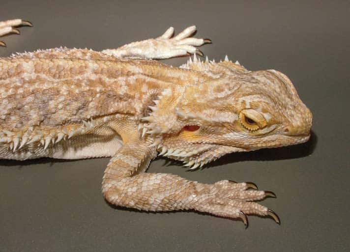
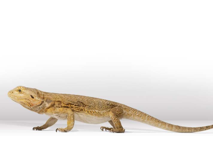
Bearded dragons are commonly presented for this problem. There are numerous potentional causes, including spinal and pelvic deformities that are secondary to nutritional secondary hyperparathyroidism (more on this later), insufficient fiber, parasites, eggs, and sometimes masses.
eric isselee/shutterstock
Bearded dragons do not drink from water bowls, and if they are not misted regularly may become dehydrated.
Bearded dragons often ingest their bedding when they are eating, and bedding such as sand, crushed walnuts, sani chips and coconut can block their intestines.
stephen barten, dvm
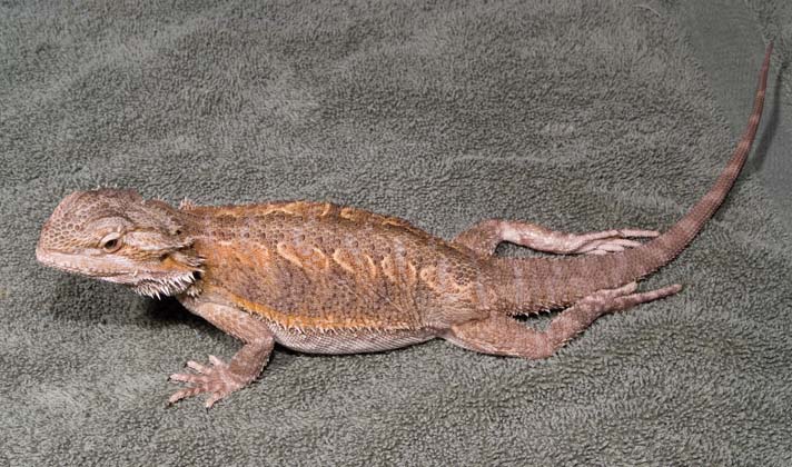
This bearded dragon has severe metabolic bone disease.
In addition, bearded dragons do not commonly drink from water bowls, and if they’re not misted regularly with water, this can lead to dehydration. Bearded dragons absorb water from the colon to keep themselves hydrated, and dehydration can lead to the ingesta becoming very dry and hard, thus obstructing the lizard’s digestive tract.
Clinical signs
- Refusal to eat
- Lack of defecation
- Straining to defecate
- Lack of energy
Diagnostics
- A physical exam will reveal a firm, oblong mass in the bearded dragon’s colon, causing pain to the lizard.
- X-rays (radiographs) can often show a distention in the colon.
- Radiographs with contrast (barium) can evaluate the colon and be used to evaluate the treatments being used.
- A complete blood panel can provide information about the overall health of the lizard and possibly uncover hidden illness.
Treatment
- Rehydration is the most important factor in treating constipation/obstipation in bearded dragons. Fluids can be given orally, by soaking, or via stomach tube and/or injections. Soaking the lizard in a shallow, warm-water bath for 30 minutes daily can be very helpful.
- Oral administration of olive oil is often recommended.
- The use of oral osmotic diuretics (Lactulose) over several days is sometimes recommended.
- Severe cases that do not respond to the previous treatments may require surgical intervention.
Prevention
- Identifying and treating any underlying medical cause is mandatory.
- Correcting any dietary and environmental errors is crucial.
Nutritional Secondary Hyperparathyroidism (NSHP)
NSHP is a form of metabolic bone disease. It is most commonly caused by owners who don’t feed their bearded dragons supplemented insects. A diet that is too high in fruit can contribute to this condition, as well.
Clinical signs
- Weight loss
- Refusal to eat
- Trembling
- Weakness
- Bone fractures (in severe cases)
- Soft jaw
Diagnostics
- A complete blood panel, including a test for ionized calcium
- X-rays
- A thorough history and physical exam
Treatment
- Nutritional support via stomach tube or assist feeding twice daily
- Calcium supplementation, orally or via injections
- Vitamin D
Prevention
- Proper diet
- 1/4 Tums tablet per kilo of body weight, ground into powder and mixed with salad daily
- Supplemented prey items
Adenovirus
This is a widespread virus that can remain non clinical in bearded dragons. It affects mostly young dragons.
Clinical signs
- Anorexia
- Failure to thrive
- Diarrhea
- Shaking
- Weakness
- Weight loss
- Head tilt
- Paralysis
- Acute death
Diagnostics
- Clinical signs and failure to find another cause for these clinical signs can indicate this virus.
- Complete blood sample
- PCR; this is a special test done at a lab using a sample taken from the lizard’s cloaca.
- Necropsy ( autopsy) of certain organs
Treatment
- Currently, there are no treatments available for adenovirus.
- Supportive care, but most lizards die once they become sick.
Prevention
- Screen all new bearded dragons coming into an existing collection.
Parasites
The two most common parasites that affect bearded dragons are coccidia (Isospora amphiboluri and Eimeria pogonae) and pinworms. Isospora species cause more severe disease.
Most lizards found in nature have these parasites in small numbers, and they do not cause a problem. The stressors of captivity, however, can sometimes create an imbalance with these parasites leading to significant illness or even death.
Clinical signs
- Lethargy
- Anorexia
- Diarrhea
- Weight loss
- Impaction
- Cloacitis
Diagnostics
- Fecal direct and float
Treatment
- Ponazuril rapidly eliminates coccidia.
- Enclosure cleaning with disinfectants and good hygiene is crucial.
Prevention
- Good hygiene
- Screening of fecal samples when bringing new lizards into a collection
Yellow Fungus Disease
(Chrysosporium anamorph)
Veterinarians are seeing this disease — a type of fungus that has been affecting various reptile species — with increasing frequency.
Clinical signs
- Yellow lesions on the top and bottom of the skin, which can become necrotic and progress to deeper lesions
- Excess scaling of the skin
Diagnostics
- Physical exam findings
- PCR testing
Treatment
- Topical and oral anti-fungal agents
Prevention
- Screen incoming lizards before adding them to an existing collection
Obesity
Obesity is common in bearded dragons. Overfeeding and lack of brumation can cause this condition, as well as feeding fruits high in sugar and larval insects high in fat. Obesity can eventually lead to a serious condition called hepatic lipidosis which can be life threatening.
Clinical signs
- Lack of energy
- Loss of interest in food items once hepatic lipidosis occurs
- Will only eat if food item is put directly in front of the lizard
Diagnostics
- A physical exam will reveal oversized coelomic fat pads.
Treatment
- Feeding a balanced diet with plenty of greens
- Increase exercise outside the enclosure
Green Iguana
Green iguanas are herbivorous (eat vegetables and plants only) from birth. Some people think they start out eating insects and later in life eat vegetables and plants. This is a misconception and, along with other misconceptions, leads to improper feeding and care for this species.
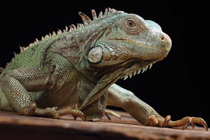
v.gi/shutterstock
Green iguanas are herbivores and should not be fed protein.
Metabolic Bone Disease
This is a complex disease that could be covered in an entire article on its own (and has, in past issues of REPTILES). To keep it simple, we will consider it to be caused most commonly by a lack of appropriate diet and natural sunshine.
Clinical signs
- Stunted growth
- Cessation of walking
- “Popeye” arms and legs. This is caused by fibrous material being laid down because calcium is removed from the bones.
- Rubbery lower jaw
- Unable to eat
Diagnostics
- Complete physical exam
- Complete blood count and chemistry
- Ionized calcium level
- X-rays
Treatment
- Supplementing calcium orally and, sometimes, by injection
- Exposure to natural sunshine
- Fluids orally
- Splints (if bones are broken)
- Feeding appropriate greens and supplements
Prevention
- Feeding appropriate diet, especially when iguanas are young
- Exposure to unfiltered sunshine
- Calcium supplementation
Constipation
Constipation is commonly caused by dehydration and lack of a proper diet, including one that is too low in fiber.
Clinical signs
- Not eating
- No defecation
- Straining to defecate
- Lack of energy
Diagnostics
- Palpation of firm stool in colon
- Radiographs can reveal large amounts of stool in colon.
Treatment
- Enemas
- Soaking
- Increase fiber in diet
- Sometime laxatives such as lactulose are used
- Improving husbandry
Prevention
- Proper fiber in diet
- Proper humidity of 80 to 90% in environment to prevent dehydration
- Exposure to natural, unfiltered sunshine
Dystocia (egg binding)
This is a common disease in females iguanas and is usually a multi-factorial problem that can be prevented.
All female iguanas can produce eggs without the presence of a male. Females that have eggs become distended and stop eating for up to four weeks. They continue to be bright, alert and active, but they may become sick when the eggs do not go through their normal cycle.
Causes of dystocia may include cool temperatures, malnutrition, dehydration and lack of a suitable substrate in which the female can lay the eggs. In addition, anatomical defects can cause a green iguana to not to lay her eggs. Problems that can cause obstruction include enlarged kidneys, abscesses, and bladder stones.
Clinical signs
- h Anorexia
- h Lethargy
- h Weakness
- h Abdominal distention
Diagnostics
- Palpation
- X-rays
- Ultrasound
Treatment
- Calcium supplementation
- Proper heat and humidity
- In some cases, spaying is required to remove the eggs and prevent further egg production.
Prevention
- Proper substrate for laying eggs
- Proper heat and humidity
- Proper calcium supplementation
- Spaying
- Exposure to unfiltered sunshine
Urinary Calculi
This condition — stones forming in the urinary tract — is being seen more commonly in iguanas. Nobody knows the underlying cause, but some suggest a lack of proper humidity, leading to chronic dehydration, can contribute.
Clinical signs
- Anorexia
- Straining to urinate or defecate
- Blood in urates
- One or two-sided rear limb paralysis (sometimes)
Diagnostics
- Can sometimes palpate the stone if it’s large enough
- X-rays
- Ultrasound
Treatment
- Surgery is the only way to remove urinary calculi. They are usually too large to pass on their own.
Prevention
- Proper heat and humidity
- Proper diet
- Frequent soaking; hydration
- Annual veterinary visits
Thermal Burns
Thermal burns result due to improper use of heat lamps, “hot rocks” or under-tank heating systems. Green iguanas are not smart enough to keep a safe distance from a heat lamp, and will climb to the highest spot to get the most heat. If the heat lamp is closer than 14 inches, a burn can result. “Hot rocks” can also malfunction, causing significant burns, and are not usually recommended. Under-tank heating systems, if used incorrectly, can heat up the bottom glass of a terrarium and burn the lizard.
When using any type of heating equipment, always be sure to follow manufacturer instructions.
Clinical signs
- Anorexia
- Lethargy
- Lesions on the back, face and belly
Diagnostics
- A physical exam can reveal necrotic areas of skin.
- Cytology of skin lesions
Treatment
- Debridment of the affected areas
- Topical antibacterial ointments
- Systemic antibiotics
- Pain medications
Prevention
- Keep heat sources a minimum of 14 inches from the highest point where the green iguana can bask.
- Do not use hot rocks.
- Do not use under-tank heating sources with green iguanas.
Leopard Gecko
The leopard gecko is a very popular pet — possibly the second most popular after the bearded dragon. It is typically a hardy species that does well in captivity if kept under optimum condtions.
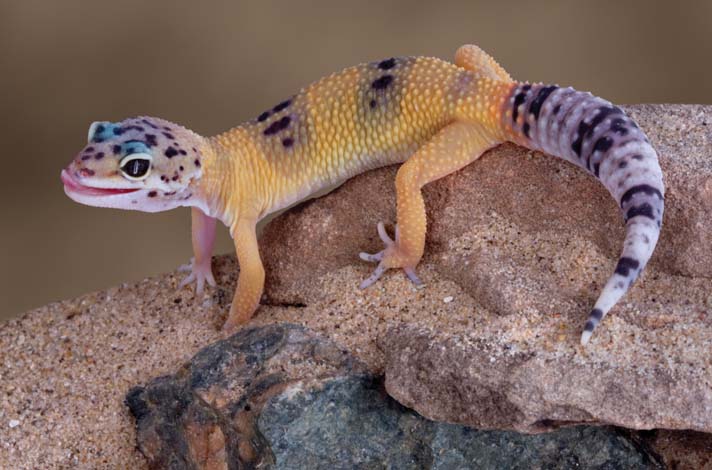
cathy keifer/shutterstock
Cryptosporidiosis can quickly kill a leopard gecko.
Hypovitaminosis A
Many reptile vitamins contain beta carotene instead of preformed vitamin A, and leopard geckos need the latter.
Clinical signs
- Anorexia
- Difficulty catching prey items
- Retained shed
- Oral lesions
- Eye lesions
Diagnostics
- Thorough history from owner regarding items being fed to the gecko
- Thorough examination
Treatment
- h Injectable vitamin A
- h Proper diet
Prevention
- Supplementation with Vitamin A (not beta carotene)
Metabolic Bone Disease
All prey items need to be supplemented with calcium before being fed to a leopard gecko. “Gut loading” crickets and mealworms will provide the best source of calcium and vitamin D.
Clinical signs
- Soft rubbery bones
- Unable to get prey
- Unable to chew prey items
Diagnostics
- Complete history
- Physical exam
- Complete blood count and chemistry
- Ionized calcium
Treatment
- Caloric supplementation
- Calcium supplementation, oral or injectable
Prevention
- Calcium supplementation of prey items
- Studies have shown exposure to unfiltered sunshine improves calcium levels. Exposure for less than 30 minutes daily has proved beneficial.
Sand Impaction
This is a common problem with leopard geckos (and other lizards) that are kept on a sand substrate.
Clinical signs
- Abdominal distention
- No feces produced
- Straining to defecate
- Anorexia
Diagnostics
- Abdominal palpation
- X-rays
- Trans illumination (the shining of a light source through the body wall to observe the intestines)
Treatment
- Oral or injectable fluid therapy
- Gastrointestinal pro kinetics (stimulate gastrointestinal tract)
- Surgery (severe cases)
- Increase fiber in the diet
Prevention
- Feed the gecko outside the enclosure, on a non-sand substrate
- Offer food in a dish
- Do not use sand substrate, even if the product packaging says it’s digestible.
- To gauge whether your leopard gecko is eating sand, mix fecal material with water and see if any sand sinks to the bottom of the glass.
Cryptosporidiosis
Caused by Cryptosporidium parasites, cryptosporidiosis can kill leopard geckos quickly. It is also known as “pencil tail” or “stick tail” due to the drastic weight loss of the gecko.
Clinical signs
- Weight loss
- Anorexia
- Regurgitating food
- Diarrhea
Diagnostics
- Autopsy of a deceased pet
- Fecal testing using a special solution
- Acid fast testing of the feces
- IFA antibody testing (a special test conducted in a laboratory that specializes in reptiles)
- DNA testing from a cloacal swab
- Other tests that are outside the scope of this article
Treatment
- Azithromycin and paramyocin. Though these medications will not “cure” your gecko, they can help control the number of parasites. Life-long use may be required.
- Supportive care, including soaking and assist feeding
- Corrective husbandry, including dietary supplementation
- Keeping the gecko hydrated using a humidity box
Prevention
- A “crypto” leopard gecko will have this disease its whole life, and must be housed separately from non-crypto geckos to avoid spreading the disease.
- Disinfect tools or other items that come in contact with the infected lizard.
Phalangeal Dysecdysis (retained shed on digits)
This condition may result in the eventual loss of digits due to circulation being cut off.
Clinical signs
- Shed is retained on the digits
- Missing digits
Diagnostics
- Careful examination with magnification will reveal multiple layers of retained shed.
Treatment
- A moist humidity box for shedding
- Soaking the toes in warm water and gently removing the retained layers of shed
- In severe cases, amputation of the affected digits may be required.
Prevention
- Leopard geckos should always have access to a humidity chamber. This can be constructed using a small plastic container with a lid, with an entry hole cut into the lid. Fill the container with moistened sphagnum moss, and keep it moist (but not dripping wet).
Crested Gecko
The crested gecko, also known as the eyelash geckos due to the elongated scales over the eyes, has become a very popular lizard pet over the past several years. It may not yet rival the bearded dragon or leopard gecko, but with increasaed captive-breeding efforts, many different types of crested gecko are now available.
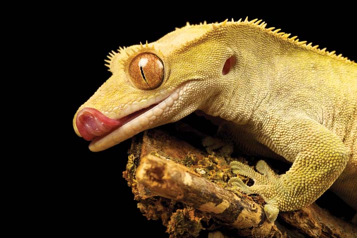
mark bridger/shutterstock
A retained shed in crested geckos (and other lizards) is usually a sign that the humidity in the enclosure is not high enough. Provide a “humidity chamber” to help prevent stuck sheds.
Metabolic Bone Disease
Clinical signs
- Stunted growth
- Gecko no longer walks
- “Popeye” arms and legs, caused by fibrous material being laid down because calcium is removed from the bones
- Rubbery lower jaw
- Unable to eat
Diagnostics
- Complete physical exam
- Complete blood count and chemistry
- Ionized calcium level
- X-rays
Treatment
- Supplementing calcium orally, and sometimes via injection
- Exposure to natural sunshine
- Fluids orally
- Splints (if bones are broken)
- Feeding appropriate insects, fruits, nectar and supplements
- Removal of branches that the gecko could fall from, causing further injury
Prevention
- Feeding appropriate diet, especially when young
- Calcium supplementation
Egg Binding
Female crested geckos may sometimes experience difficulty passing eggs, either due to physiological problems or a lack of a proper egg-laying substrate.
Clinical signs
- Anorexia
- Lethargy
- Weakness
- Abdominal distention
Diagnostics
- Palpation
- X-rays
- Ultrasound
Treatment
- Calcium supplementation
- Proper substrate for laying eggs
- Proper heat and humidity
- Medications to help stimulate egg laying (oxytocin). While no studies have proven this to be an effective treatment, I have found it to be helpful in certain cases.
- Surgical intervention (last resort)
Tail Injury
Tail injuries are common with crested geckos, and they do not regrow their tails. A stub will result.
Clinical signs
- Damage to the tail
Diagnostics
- Evaluate if the tail is viable or not.
Treatment
- Amputation, if indicated
- Application of topical antibacterial
- Oral antibacterial
- Pain medications
Sand Impaction
This is a common problem with lizards kept on sand.
Clinical signs
- Abdominal distention
- No feces produced
- Straining to defecate
- Anorexia
Diagnostics
- Abdominal palpation
- X-rays
- Trans illumination (shining a light source through the body wall to observe the intestines)
Treatment
- Oral or injectable fluid therapy
- Gastrointestinal pro kinetics (stimulate gastrointestinal tract)
- In severe cases, surgery may be needed.
- Increase fiber in the diet
Prevention
- Feed the gecko outside the enclosure, on a non-sand substrate
- Offer food in a dish
- Do not use sand substrate, even if the product packaging says it’s digestible.
- To gauge whether your crested gecko is eating sand, mix fecal material with water and see if any sand sinks to the bottom of the glass.
Retained Shed
Clinical signs
- Layers of retained shed on various parts of the body
Diagnostics
- Careful examination with magnification will reveal multiple layers of retained shed.
Treatment
- Provide a moist humidity box for shedding.
- Soak the affected areas in warm water and gently remove the retained layers of shed.
Prevention
- As mentioned for leopard geckos, provide a humidity chamber using a small plastic container with a lid, with an entry hole cut into the lid. Fill the container with moistened sphagnum moss, and keep it moist (but not dripping wet).
Green Anole
Long a staple in the pet reptile industry, the green anole remains a favorite pet lizard. Unfortunately, due to their inexpensive price, green anoles are often bought on impulse and do not always receive the proper care, leading to early death.
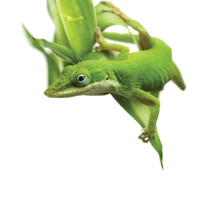
leigh prather/shutterstock
Green anoles may exhibit trauma, which can result from two males fighting. It’s not a good idea to keep multiple males together.
Sand Impaction
This is a common problem with lizards kept on sand.
Clinical signs
- Abdominal distention
- No feces produced
- Straining to defecate
- Anorexia
Diagnostics
- Abdominal palpation
- X-rays
- Trans illumination (shining a light source through the body wall to observe the intestines)
Treatment
- Oral or injectable fluid therapy
- Gastrointestinal pro kinetics (stimulate gastrointestinal tract)
- In severe cases, surgery may be needed.
- Increase fiber in the diet
Prevention
- Feed the anole outside the enclosure, on a non-sand substrate
- Offer food in a dish
- Do not use sand substrate, even if the product packaging says it’s digestible.
- To gauge whether your crested gecko is eating sand, mix fecal material with water and see if any sand sinks to the bottom of the glass.
Metabolic Bone Disease
Clinical signs
- Stunted growth
- Anole no longer walks
- “Popeye” arms and legs, caused by fibrous material being laid down because calcium is removed from the bones
- Rubbery lower jaw
- Unable to eat
Diagnostics
- Complete physical exam
- Complete blood count and chemistry
- Ionized calcium level
- X-rays
Treatment
- Supplementing calcium orally, and sometimes via injection
- Exposure to natural sunshine
- Fluids orally
- Splints (if bones are broken)
- Feeding crickets, mealworms, fruit flies, Phoenixworms. All these should be gut loaded and dusted prior to feeding.
Prevention
- Feeding appropriate diet, especially when young
- Calcium supplementation
Trauma
Trauma in green anoles is most commonly caused by males fighting or aggressive handling by owners.
Clinical signs (can vary depending on type of injury)
- Broken tails
- Ripped skin
- Broken bones
- Bite wounds
- Color change from green to brown is a sign of severe stress
Diagnostics
- Thorough physical exam and history
Treatment
- Housing males separately
- Oral and topical antibiotics
- Pain medications
Prevention
- Do not keep male anoles together
- Practice gentle handling, if at all
- Provide proper diet and environment
Parasites
It’s common for a green anole to be heavily parasitized, and the most common parasites that affect anoles are coccidia (Eimeria spp.). In addition, helminths and cestodes have been found. Most wild anoles have these parasites in small numbers, and they do not cause a problem for the lizards. The stressors of captivity, however, can sometimes create a parasite imbalance, causing significant illness or even death.
Clinical signs
- Lethargy
- Anorexia
- Diarrhea
- Weight loss
Diagnostics
- Fecal direct and float
Treatment
- Ponazuril rapidly eliminates coccidia.
- Environmental cleaning with disinfectants and good hygiene is important.
- Depending on the parasite found, other dewormers may be used.
Prevention
- Good hygiene
- Screening of fecal samples when bringing new lizards into a collection.
Foreign Body
Green anoles will occasionally ingest abnormal items, leading to obstruction.
Clinical signs
- Anorexia
- Regurgitation of food
- Weight loss
Diagnostics
- X-rays
- X-rays with contrast
- Trans illumination (shining a light source through the body wall to observe the intestines)
- Ultrasound
Treatment
- Oral fluids
- Injectable fluids
- High fiber
- Surgery (in severe cases)
Prevention
- Do not put anything that your green anole could ingest in the cage that is not digestible, including substrate comprised of particles your pet may be tempted to eat.
It’s important to understand that all of the conditions mentioned in this article are preventable as long as lizard owners provide the proper care, including diet, that is appropriate for the species of reptile that they are keeping. Remember, too, that not all information found on the internet is accurate in regard to keeping and feeding reptiles.
Being a responsible reptile owner includes finding a reptile veterinarian in your area and getting professional advice on how to care for your reptile. You can find a list of reptile veterinarians at the Association of Reptile and Amphibian Veterinarians website at arav.org.
Tia Greenberg, DVM, completed her schooling at Ross University in 1992. After completing a year at The Animal Medical Center in New York City, she accepted an associate position working with Dr. Douglas Mader to improve her knowledge and skills in reptile medicine and surgery. She is currently the chief of Medicine and Surgery at the Westminster Veterinary Group in Southern California (westminsterveterinarygroup.com). She has been practicing reptile medicine for over 25 years and is a member of the Association of Reptile and Amphibian Veterinarians.
