Reptile eye problems and their symptoms.
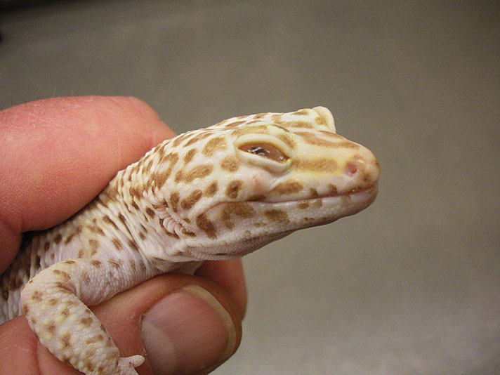
“I don’t think Spot can see,” the little girl answered when I asked her why she brought her beloved leopard gecko in for an exam. “He used to chase crickets around the cage, now he ignores them, and he’s getting skinny.” Sure enough, both eyes were sealed shut. Now, we had to figure out why.
tom greek
There is a small abscess below the eye of this leopard gecko. Note the dried debris collecting in the eye.
Some poets may say eyes are the windows to the soul, but veterinarians think of eyes as the windows to the health of an animal. Even diseases from other parts of the body may show ocular symptoms, not just primary eye diseases. It is easy to focus on the primary problem and miss others, so it is extremely important to perform a complete physical exam on any sick animal.
When performing a physical exam, you must know what is normal before you can spot the abnormal. Therefore, it is highly recommended to do physicals on your pets regularly. There will be variations between species as well as individuals, so the more you practice, the better you will become.
Magnification is essential for ophthalmic examinations. I’ve got a fancy ophthalmoscope with its own light source and varying amounts of magnification. Veterinary ophthalmologists have even better equipment, but unless it’s a tool you are going to be using every day, there is no need to spend hundreds or thousands of dollars on special equipment. A good magnifying glass and a bright desk lamp should be enough for most of hobbyists.
Reptile Eye Anatomy
tom greek
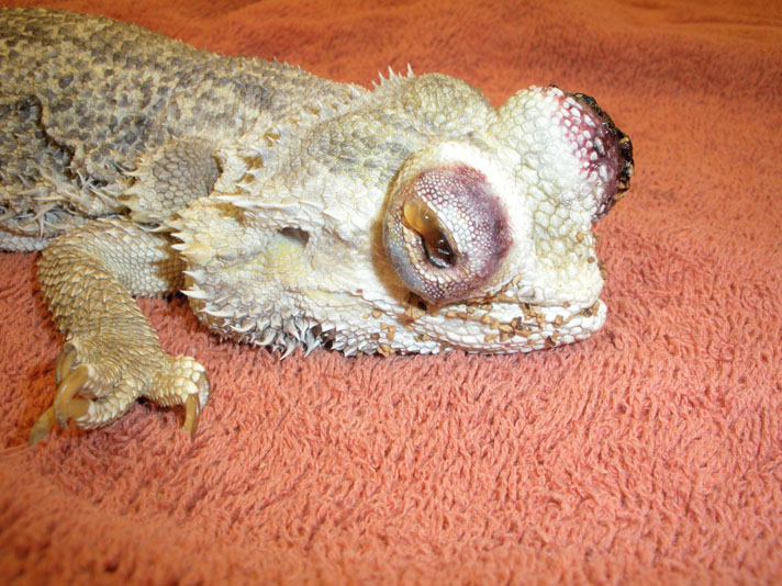
Crushed walnut shell bedding caused severe damage to the eyes of this bearded dragon.
All reptiles have eyelids. For most lizards, turtles and tortoises, it is the lower lid that is moved more, while the upper lid is more mobile in crocodiles and alligators. Some species have very thin lids, with few scales, allowing some vision even when the eyes are closed. Most reptiles with movable eyelids also have a third eyelid underneath the upper and lower lids.
All snakes and some lizards have a fused, immobile lid covering the eyes, called a spectacle. Many think that a snake’s eyes are always open. In fact, their eyes are always closed. Look closely and you will see the eyes moving freely underneath the clear spectacle.
The conjunctiva is the tissue lining the inner lids to the sclera or eyeballs itself. The clear surface of the globe is called the cornea. The iris is the colored portion inside the eye. By opening and closing, it allows light through the hole called the pupil onto the light receptors or retina on the back surface of the eye. Some species have round pupils, others elliptical openings. The shape of the pupil can give a clue to the diurnal or nocturnal activity patterns in a reptile. Species with round pupils tend to be active during the day while those with slit pupils tend to be active at night. Some nocturnal species of geckos have tiny indentations along the pupil margin. In bright light, when the pupil would be only a thin slit, the small holes create multiple tiny openings, allowing better vision than the slit alone would afford. The eastern box turtle shows sexual dimorphism with the iris color —males tends to have red irises while females have brown. The lens behind the pupil focuses the light onto the retina.
Diseases of the Eye in Reptiles
Keep an eye out for these problems.
Eye Discharge in Reptiles
tom greek
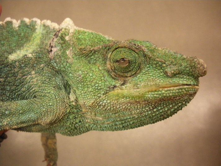
Not only is there ocular discharge, but thick mucus in this chameleon’s mouth shows how systemic problems may sometimes present as eye symptoms.
The eyes are kept moist and clean by a constant supply of tears. If there is something irritating the eye, there may be an overproduction of tears, or the tears may change consistency.
The eye has a drainage system called the nasolacrimal duct that brings the tears to the nasal canal. Think about when your eyes water—your nose runs. If this duct is blocked, the tears have no place to go and will run down the face. For species with a spectacle that develop a blocked nasolacrimal duct, the tears will build up, thus creating a bulging spectacle. This requires surgery.
Some species of tortoises, such as Aldabra and red-footed tortoises, will exhibit a normal, mild, clear, ocular discharge, although it is thought that inadequate humidity may play a part in this.
Retained Shed in Reptiles
tom greek
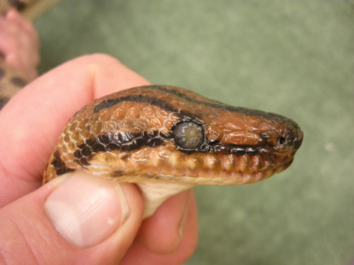
Damage to a python’s spectacle, from an aggressive attempt by the owner to remove a retained shed.
The primary cause for retained shed is husbandry problems, most often inadequate humidity. But other causes, such as wound on the lids or snake mites collecting along the spectacle margin, may prevent proper shedding.
If this occurs, try to gently remove the retained shed as soon as it is noticed. The longer you wait, the harder it can be to remove. Be gentle when removing a retained shed on the spectacle. Some people like to use moistened cotton swabs, others use a little piece of cellophane tape. If it does not come off easily—STOP! You may do more harm than good if you continue. It is time to seek professional help. I’ve seen numerous cases of permanent damage with over-aggressive, but well-meaning, individuals ripping off spectacles.
Shed collecting along the inside of the lids is a common problem for leopard geckos and chameleons. I use saline to flush the eyes, a pair of very tiny forceps to help remove the shed and big magnifying loupes to see what I’m doing. Again, if you don’t feel comfortable poking around your lizards eyes, then you probably shouldn’t be doing it. Some species, such as ball pythons, may have a normal dent or wrinkle on their spectacles—do not mistake this as a retained spectacle.
Reptile Eye Trauma
tom greek
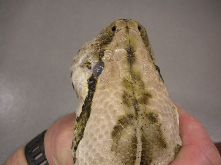
A bite from a live rat caused an infection below this boa’s eye, causing fluid to build up in the subspectacular space.
Trauma may be mild abrasions from rubbing on cage surfaces to lacerations that may heal on their own or with topical antibiotics, to more severe lid lacerations requiring surgical closure for proper healing. Bite wounds from live prey can be deep enough to destroy the globe, requiring enucleation or removal of the eye.
Foreign Bodies in Eyes of Reptiles
tom greek
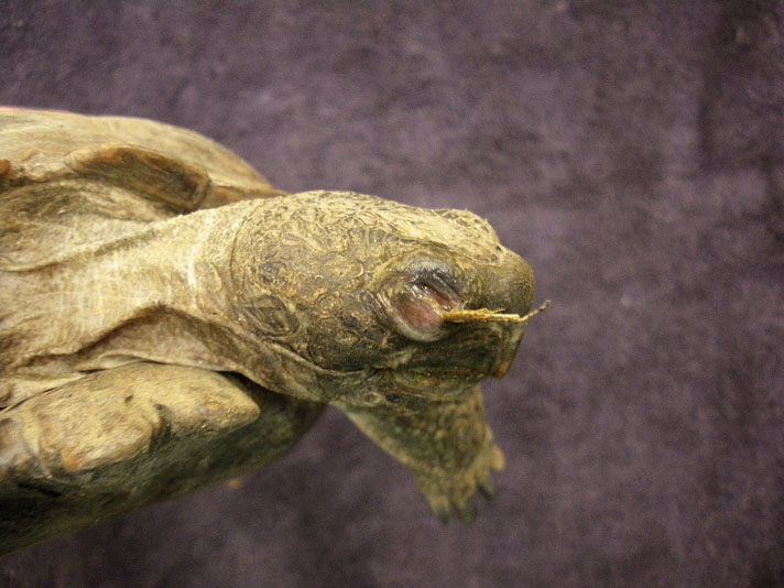
A grass awn (foxtail) in the eye of a desert tortoise.
I’ve seen a few cases of grass awns getting stuck in the conjunctival tissue under the eyelids of lizards or tortoises housed outdoors. More common, though, is bedding material collecting in the eyes. Sand, bark dust, coconut fiber—if it is small enough to get in the eyes, I’ve probably seen it. Outward symptoms may include ocular discharge, blepherospasm (squinting) or corneal haziness. Treatment involves removing the offending material and topical medication to help healing.
Conjunctivitis in Reptiles
This is a term used for inflammation of the conjunctiva. The classic presentation is the red-eared slider with puffy eyes. There can be a couple of causes. Poor water quality can lead to a bacterial conjunctivitis. Similar in appearance is a vitamin A deficiency. Given that it usually takes a biopsy to tell the difference, I will often treat for both with vitamin A supplementation and topical antibiotics.
We usually think of aquatic chelonians as having vitamin-A deficiencies. There is a theory that some of the eye problems we see in leopard geckos and chameleons may also be vitamin-A related. Further research is needed, as overdosing vitamin A can be toxic.
Conjunctivitis in snakes may present itself as abscess formation under the spectacles. Trauma and local and systemic infection can also cause subspectacular abscesses. Surgery is usually required to remove the purulent material.
Corneal Lesions in Reptiles
tom greek
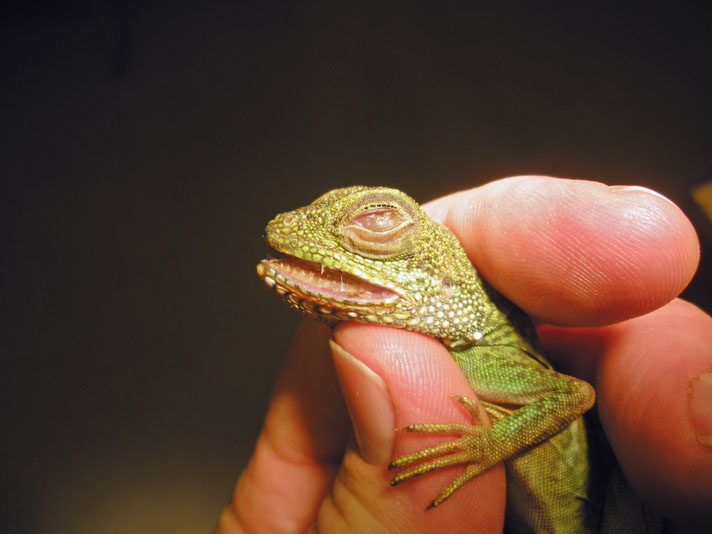
The cornea should be smooth and clear, not opaque and rough as in this water dragon.
A healthy cornea is clear. Any change in color may be a sign of a problem. As the cornea swells, it may become hazy or opaque. We use a topical fluorescein dye and a black light to identify scratches or ulcers on the surface of the cornea. Chronic irritation may cause pigmentation to the cornea. Treatment for corneal damage is usually topical medication to prevent infection and promote healing. Occasionally, surgery is required.
Anterior Uveitis in Reptiles
The anterior chamber is a fluid-filled space under the cornea. Blood or pus in the anterior chamber can be a sign of local trauma or infections. It can also be a sign of septicemia or infection spread throughout the entire body.
Reptile Cataracts
Behind the iris is the lens. The job of the lens is to focus the light beam on the light-sensing nerves of the retina. With age, the lens may lose some of its elasticity and lose some ability to focus. This is why many people require reading glasses as they get older. Also with age, as well as some systemic diseases, the lens may become opaque, causing what we know as cataracts. This can lead to blindness, but cataract surgery is available.
Congenital Abnormalities in Reptiles
Occasionally, hatchlings will be born with eye problems, the most common being microphthalmia. The eyes appear very small or nonexistent (there is usually a microscopic eye remnant present). In the wild, these cases would carry a very poor prognosis of survival, but I’m surprised how well many of these babies do in captivity.
tom greek
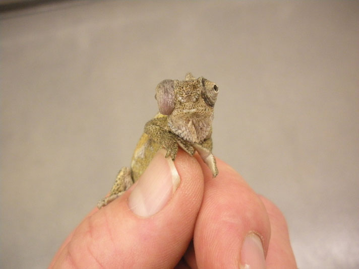
Infection in the eye of a chameleon.
Some systemic diseases may appear to be ocular diseases. Calcium deficiencies can lead to malformation of the skull, causing a bug-eyed look with the eyes not fitting properly into the eye sockets. While we can treat the underlying disease, the skull deformity may be permanent. Eyes that don’t fit properly may also have trouble blinking, leading to a portion of the cornea to dry out and become irritated.
Other causes for exophthalmia (bulging eyes) include retrobulbar abscesses, tumors, vascular obstruction and edema. Again, rarely is it the eye itself that is having problems, but the structures around the eye that are causing it to be pushed outward.
At the opposite end of the spectrum are sunken eyes or enophthalmia. This can be a symptom of dehydration, weight loss, septicemia or impending death. A complete physical exam, as well as other diagnostic tests, may be necessary to determine the exact cause.
Chameleons, leopard geckos and water dragons are the most common species I see with eye problems. With chameleons, a lot of it has to do with their unique eyelid anatomy. Their lids are essentially fused with only a small opening. There is a theory that, in captivity, we don’t really rain on them, only mist them down, and a heavier shower would allow them to flush out their eyes. I have had many cases where all I’ve needed to do was flush out the eyes with saline and the chameleon was fine afterwards.
With leopard geckos, the problems often appear to be attributed to low humidity and poor shedding. The retained shed along the inside of the eyelids creates an irritation, which causes the eyes to remain closed as mucus and debris builds up under the lids. Unfortunately, many owners don’t notice this happening until the gecko has stopped eating and becomes thin. Sometimes the damage to the eyes is so severe that they lose their vision. Treatment involves removing the debris and using a topical medication to help the eye heal.
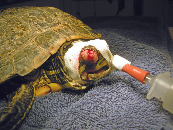
tom greek
A mass on the eyelid of a red-eared slider before removal.
Water dragons are often brought in with closed eyes. The most common cause is bedding material collecting under the lids. Many owners use coconut husk or fir bark bedding for their dragons because it will hold moisture well and help with cage humidity. But if these beddings dry out, they can be quite dusty and this dust collects in the eyes. Keep bedding moist to prevent this. Flushing the eyes with saline and topical antibiotics are used for treatment.
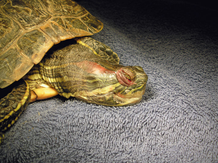
tom greek
The mass removed from the eyelid of a red-eared slider.
A Happy Ending
So how did Spot the leopard gecko do? The physical exam showed that a few of his toes had retained shed remaining on them. This is a strong clue that the humidity in his cage wasn’t adequate and most likely the cause of his eye problems, as well. The shed was removed from his toes along with the shed, dried mucus and debris from under his eyelids. Topical antibiotics were given to help the eyes heal, as well as instructions to provide a moist hide box for Spot to help with future shedding. On his two-week recheck, the eyes were open and he was back to chasing crickets.
Tom Greek, DVM, is a graduate of the University of Wisconsin, Madison, and a Southern California native. His interest in reptiles began at the early age of 5 when he received his first pet snake. He currently practices small- and exotic-animal medicine at Greek & Associates Veterinary Hospital in Yorba Linda, Calif.


