What if my snake's spectacle is retained when it shed's its skin?
"And immediately there fell from his eyes as it had been scales; and he received sight forthwith."-Acts of the Apostles, Chapter 9
That snakes lack eyelids is common knowledge. In place of the missing eyelids are protective, clear scales called the spectacles or eye caps. These scales serve the same function as eyelids: They keep dirt and debris off the cornea (surface of the eye) and prevent the cornea from drying out by holding a layer of tears in place.
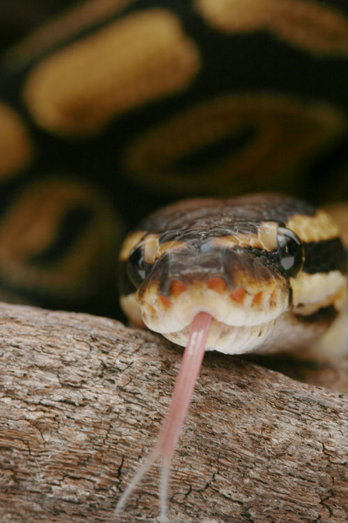
Photo by Gina Cioli/i5 Studio
Ball pythons (Python regius) are known to suffer from retained spectacles due to their bulging eyes.
The outer layer of the spectacle normally is shed along with the rest of the skin. However, sometimes that outer layer of spectacle is retained, and it fails to slough with the skin. Judging by the number of calls concerning this issue I receive from snake keepers and veterinarians alike, it is widely known that retained spectacles occur. Yet those same calls reveal that there is a lot of confusion about how to recognize retained spectacles and what to do about them. Several conditions mimic retained spectacles, and an accurate diagnosis must be made prior to treatment. Fortunately, the diagnosis is made by simple visual inspection.
Anatomy and Physiology of the Skin and Spectacles of Snakes
Reptile epidermis (the outer layer of skin) consists of two layers. The inner stratum germanitivum is the germinal layer, the cells of which divide to form new layers of skin. The outer stratum corneum is the cornified layer, consisting of a protective layer of dead, flat, horny cells filled with keratin. As cells move outward from the first layer, they gradually become the cornified cells of the second layer.
Snake skin is normally in a resting stage. The shed cycle begins with the proliferation of the basal cells of the stratum germanitivum. These cells move outward and form a new layer deep to the old stratum corneum. A cleavage zone develops between the newly generated layer and the stratum corneum. Blood and nerve supplies to the outer layer are lost, while the surface of the newly generated layer becomes cornified. Lymph fluid diffuses into the cleavage zone causing a loosening of the outer layer.
A combination of this lymph and the thickness of the newly generated layer of cells cause a temporary dulling of the color of the skin. The same mechanisms cause the normally clear spectacle to become bluish white, which is why snakes in the pre-shed phase of this cycle are said to be "opaque." The lymph between layers is resorbed, clearing the spectacles, three to five days before the snake sheds.
The normal process of shedding is important in cases of retained spectacles because it explains what lies under the retained spectacle: a normal, full-thickness spectacle consisting of both layers of epidermis, dermis, blood vessels and nerves. The cornea lies under the spectacle and is separated from it by a minute intraconjunctival space filled with tears.
Older books and articles on reptile medicine claim that irreparable damage to the cornea surely results when a retained spectacle is removed with excessive force or without prior moistening with water. This claim has some basis but is incomplete in its explanation. Obviously, a full thickness, living spectacle lies between the retained one and the cornea, so the only way for the cornea to be damaged would be for the living spectacle to be removed along with the retained one. In that horrible instance, immediate damage could result from direct contact between any instruments used and the cornea. However, a more drastic consequence is that removal of the living spectacle would be the equivalent of surgical excision of the eyelids in a person. Nothing would remain to hold or spread a protective layer of tears across the cornea, which would dry out, ulcerate, thicken with scar tissue and result in blindness, or even loss of the eye. It would take some force to traumatically rip away a living spectacle, as it would be similar to ripping off a full thickness patch of skin from the body. Nevertheless, keepers and inexperienced veterinarians occasionally do just this. A light and gentle touch when working around the eye is essential.
Snake scales are not true scales as seen in fish. Anyone who has cleaned a fish has seen individual scales detach. Snake scales are merely thickened areas of skin. There is thinner skin between, but continuous with, every so-called scale. The thin skin between scales becomes obvious when a snake eats a large meal and the skin is distended. This also is the situation with the spectacle of the eye. There is a deep periocular (around the eye) crevice. The thin skin that lines that crevice is connected to the spectacle all the way around it. When a snake sheds, it sheds this thin skin along with the spectacle, normally all connected in one piece. This is important because when a spectacle is retained, the skin being shed tears at its weakest point-through this thin skin of the periocular crevice. This leaves behind a tag of loose, dead skin around the periphery of a retained spectacle. It may be a narrow tag, but either this tag or the edge of the retained spectacle itself is always readily visible if the crevice is examined carefully.
When a mammal produces tears, the blinking eyelids spread them across the cornea. Flowing tears are collected in the nasolacrimal (tear) ducts that drain into the sinuses. Thus, when a person cries, the tears drain into the nasal cavity and he has to blow his nose. When the nasolacrimal ducts are overloaded, tears overflow and run down the cheeks.
Snakes don't cry, but do produce tears to moisturize and lubricate the cornea. Tears flow in the minute intraconjunctival space between the cornea and the spectacle. Nasolacrimal ducts drain the tears from that space, but unlike people, drain into the mouth rather than the nose. In snakes, the nostrils are located dorsolaterally at the front of the snout. The left and right internal nostrils empty into a crevice called the choana in the roof of the mouth, rather than the back of the throat. Furthermore, snakes possess paired vomeronasal organs (Jacobson's organ), which are the chemosensory organs to which the forked tongue brings scent particles. The left and right vomeronasal ducts also open on the roof of the mouth and appear as tiny slits just off the midline in front of the choana. The nasolacrimal ducts open into these vomeronasal ducts. Thus, snake tears drain into the roof of the mouth rather than into the sinuses.
The nasolacrimal duct system is important because of the spectacle. If anything blocks one or both nasolacrimal ducts, such as a mouth infection or tumor near the vomeronasal ducts, the tears have nowhere to go. The spectacle prevents them from running down the cheek as in people. Instead, the space between the spectacle and cornea distends with fluid. Sometimes the fluid is clear, while in other cases it is infected and cloudy. Occasionally people mistake the distended spectacle for a retained spectacle.
Retained Enecap in Specific Snakes
Any snake may suffer a retained spectacle, but it is most common in snakes with large, protruding eyes. Among pet snakes, ball pythons (Python regius) and indigo snakes (Drymarchon corais) are commonly affected. It is also seen in snakes with relatively small eyes, such as kingsnakes (Lampropeltis spp.).
The history often includes conditions of low humidity, such as when furnace heat is employed during winter. Low humidity may be evidenced by dysecdysis (difficult shed), where the old skin comes off in patches instead of one continuous piece. Another factor may be the presence of snake mites (Ophionyssus natricis), which often inhabit the periocular crevice causing inflammation and weakening the stratum corneum.
Infection of the skin around the eye also can predispose a snake to suffering retained spectacles. One would expect this condition to be seen only in snakes that have recently shed. While that is often the case, some owners fail to notice the retained spectacle until some time after the shed, or after a number of successive sheds result in several layers of retained spectacles.
Retained Eyecap Diagnosis
An abnormal appearance to the spectacle does not mean that a retained spectacle is present. Snakes with large, protruding eyes often develop wrinkles and creases in the spectacle. This is normal and requires no treatment. The wrinkles may become more pronounced with age.
When a snake sheds, the cast skin should be inspected for the presence of both spectacles. If one or both are missing, they may be retained and the snake should be examined. This is not a sure sign of retained spectacles as the missing ones may be found separately on the floor of the cage. Some retained spectacles have a cloudy look, so if the snake appears opaque immediately after its shed it should be examined. This is not a sure sign of retained spectacles either. I have seen snakes go through back-to-back shed cycles due to skin disease or wounds, in which case the snake is opaque for the next shed as soon as the first skin is cast. Sometimes the retained spectacles are clear, and in other individuals they are cloudy or wrinkled. If one eye looks different than the other immediately after a shed, the snake should receive an examination.
The only sure way to know a spectacle is retained is to see the edge of that spectacle or the tag of thin, dead skin attached to it in the periocular crevice. If there is not a retained spectacle, no edge will be visible because the normal spectacle is continuous with the thin skin around it. A good light source and firm restraint to prevent sudden movements on the part of the snake should be used when inspecting the eye. Magnification is useful in small specimens. In those cases in which the periocular crevice is narrow and difficult to visualize, a blunt probe may be inserted gently into the crevice to expose the edge of a retained eye cap. Avoid any probe with sharp points or edges, such as a round toothpick or straightened paper clip. A flat toothpick with rounded ends is relatively soft, flexible and less likely to damage tissue compared to a metal probe. The probe is gently inserted into the crevice at the front of the eye (3 o'clock if the right eye were the face of a clock, 9 o'clock for the left eye) until it stops, then slowly and gently is swept up and out of the crevice along the surface of the spectacle. If a retained spectacle is present, its edge and the tag of thin skin attached to it are obvious.
When using a probe, gentle handling and manipulation are essential to prevent injury to the delicate eye and surrounding tissues. It is possible to damage the eye doing this incorrectly. Do not apply force if the probe meets resistance. Use the utmost caution, and consult an experienced veterinarian when unsure of how to proceed.
In cases where the owner has been inattentive, several layers of retained spectacles can build up. The first retained spectacle sometimes prevents the next one from being shed during the next cycle, and so on and so on. Severe cases can resemble a thick pair of glasses distorting the eye.
If no edge is present but the eye bulges, there may be a blockage of the nasolacrimal duct. The bulging eye may be clear if the duct is only blocked, or cloudy if infection is present. There is no home remedy for these cases and consultation with a veterinarian experienced with snakes is necessary. Anesthesia and surgical drainage, with or without antibiotics, will be necessary. The details of that treatment are beyond the scope of this article but have been reported in the veterinary literature.
Retained Eyecap Treatment
The cardinal rules in treating retained spectacles are never to grasp any tissue with forceps or a hemostat and never to pull, yank or otherwise apply force. This is necessary to prevent the inadvertent removal of the living spectacle underneath.
If the snake is diagnosed within 24 hours of shedding its skin, the retained spectacle is likely to be loose and easy to remove. If more time than that has passed, the spectacle may have desiccated and become adherent to the living spectacle. In that case it should be rehydrated by applying a few drops of artificial tear solution, a number of brands of which are available without prescription from human pharmacies. The drops should be applied several times over 30 to 60 minutes. Initial attempts to remove the spectacle should use a piece of scotch tape wrapped around the index finger with the sticky side out. After blotting the spectacle dry, this is pressed gently against the eye and lifted off. Common sense tells us that excessive pressure could damage the eye. Usually this technique will remove the retained spectacle when it sticks to the tape.
Alternatively, a blunt probe (the end of a flat toothpick or one side of a blunt forceps or hemostat with caution not to grasp the spectacle) is inserted gently into the periocular crevice (as described above) and maneuvered under the rim of the retained spectacle. The prove is slowly and gently lifted to remove the spectacle, much like a spatula is used to lift a pancake from a griddle. Again, good restraint, a steady hand and a gentle touch are important to prevent accidental damage to the eye. Instruments with sharp points or edges have no place here.
If the retained spectacle seems stuck to the living one and is not easy to remove, you should use a conservative approach and do nothing other than correcting environmental conditions (see Prevention, below) and waiting for the next shed. In some cases the retained spectacle will come off without treatment during the next shed. In other cases the second spectacle is also retained, but both will come off easily within 24 hours of the shed. The snake must be inspected carefully within this time period.
If mites are present they must be controlled. Since no insecticide has ever been tested or approved for use in reptiles, there is much controversy about mite treatment. Because it is a complex subject worthy of a full article itself, I refer readers to the listed references for various opinions on the subject. If inflammation, discharge, swelling, bleeding, scabs, crusts or any sign of infection is present, home treatment is contraindicated and the keeper should consult a herpetological veterinarian.
Retained Eyecap Prevention in Snakes
In the absence of mites or infection, merely providing adequate humidity is usually enough to prevent retained eye caps in snakes. There are many species of snakes with many different humidity requirements, so while general recommendations will be made here, keepers should learn and provide the specific requirements of the species he chooses to keep.
Supplemental humidity can be provided using anything from fancy automatic misting systems to the occasional use of hand held sprayers. One thing to keep in mind is that keepers often seal their cages to hold the humidity in. This is a big mistake. While it is easier to maintain high levels of humidity in a closed container, the combination of heat, humidity and stagnant air fosters the growth of bacteria and fungi, resulting in skin and respiratory infections. Adequate ventilation is essential when providing supplemental humidity.
One simple method of providing supplemental humidity is the use of a so-called humidity box. This is a plastic container big enough to hold the snake, with an access hole cut in the lid. Plastic food storage containers and show and sweater boxes work well. The box is partially filled with damp sphagnum moss, which is available at any store which sells house plants. The moss comes dry, and is moistened by soaking in water and wringing it out like a sponge. This provides adequate, but not excessive, moisture. As time goes on the water will evaporate, and the moss needs to be remoistened on a regular basis. Sphagnum moss is naturally acidic and will discourage the growth of mold and bacteria. Nevertheless, if the humidity box contains visible mold, if it smells foul or if the snake defecates in the box, replace the moss and wash the box thoroughly. Place the box half on and half off the heat source so it contains warm and cool ends, allowing the snake to thermoregulate while seeking high humidity. An additional dry hide box should be provided so the snake can choose dry or moist conditions according to its needs, yet still enjoy visual security.
Summary
Remember the creed of the health care profession: Primum non nocere-"First, do no harm." Retained spectacles are relatively easy to treat, but there is a risk of causing permanent blindness through overzealous manipulation. At the same time, this never is a life-threatening condition requiring emergency treatment. When in doubt, it would be a wise investment to consult an experienced veterinarian and learn the proper technique.
References
Cooper, J.E. and O.F. Jackson (eds.). 1981. Diseases of the Reptilia. Academic Press: London
DeNardo, D. and E.J. Wozniak, 1997. Understanding the snake mite and current therapies for control. Proceedings, Association of Amphibian and Reptilian Veterinarians: Houston
Frye, F.L. 1991. Biomedical and Surgical Aspects of Captive Reptile Husbandry. Krieger Publishing Company: Malabar.
Klingenberg, R.J. 1993. Understanding Reptile Parasites. California Advanced Vivarium Systems: Lakeside.
Mader, D.R. 1996. Dysecdysis: abnormal shedding and retained eye caps. In: Mader, D.R. Reptile Medicine and Surgery. W.B. Saunders Company: Philadelphia
Millichamp, N.J., E.R. Jacobson and E.D. Wolf, 1983. Diseases of the eye and ocular adnexae in reptiles. Journal of the American Veterinary Medical Association, 183 (11): 1205-1212.
Porter, K.R. 1972. Herpetology. W.B. Saunders Company: Philadelphia.
Underwood, G. 1970. The Eye. In: Gans, C. and T.S. Parsons (eds.), Biology of the Reptilia, vol. 2, pp. 1-97. Academic Press, Inc.: New York.
Williams, D.L. 1996. Ophthalmology, In: Mader, D.R. Reptile Medicine and Surgery, pp. 175-185. W.B. Saunders Company: Philadelphia.