There are more than 7,000 species of amphibians, with the actual numbers changing daily (see amphibiaweb.org for the most recent data). This group of
There are more than 7,000 species of amphibians, with the actual numbers changing daily (see amphibiaweb.org for the most recent data). This group of vertebrates has been around for more than 300 million years. Although amphibians are the first vertebrates to colonize on land, they still rely on an aquatic environment for breeding and development of young. For the most part, amphibians have an aquatic larval stage followed by a terrestrial adult stage. This dual lifestyle can play an important role in understanding many of the medical problems with these animals.
Amphibians, which are members of the class Amphibia, can be broken down into three main groups, called orders: Gymnophiona (caecilians), Urodela (salamanders and newts) and the Anura (frogs and toads). With more than 7,000 species to consider, it is not possible to cover all the specific conditions unique to each one. For this article, the discussions will cover general issues surrounding each of the three groups.
Douglas mader, dvm
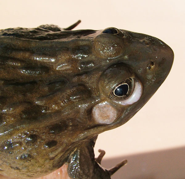
Hypovitaminosis A—note the swollen conjunctiva around the eye.
Amphibian Anatomy and Physiology
The caecilians represent the smallest group and are the least common amphibians seen in captivity, but interest in this unique order is growing and numbers in captivity are increasing.
These animals are elongated and look more similar to a worm than a frog. There are a few aquatic species, but most are burrowing terrestrial animals. The eyes are reduced in size or absent. They have skulls that are designed for burrowing and lack limbs that would normally hinder tunneling through the dirt.
Urodela, the next largest order, splits their numbers approximately in half, with half living an aquatic lifestyle and the others a terrestrial one. These animals have a generalized lizard-like body plan with a head, four limbs, long body and a tail. However, that said, many species have reduced limbs and all species have fewer toes than their lizard counterpart. There are unique characteristics in some urodelans, such as the lungless salamander, the eyeless olm and the axolotl, a species that demonstrates neotany, a condition where the adults resemble gilled larvae.
The anurans, the frogs and toads, are the largest order, with more than 6,000 species. Most of the anurans are terrestrial, but there are a few species that are entirely aquatic.
Frogs have four limbs, no neck, a broad head and a reduced or absent tail. Their rear legs are much longer and larger than the front limbs, which support their saltatorial lifestyle (hopping), but interestingly, many frogs will “walk” rather than jump.
Anurans are used commonly in biomedical research and are becoming more popular as pets.
Healthy Housing for Amphibians
Given that many amphibian maladies are related to husbandry and nutrition, a short, general discussion about caging is in order. Perhaps one of the most important differences between amphibians and the more popular reptile pets is temperature. Where herpetoculturists are required to provide appropriate warm temperatures, thermal gradients and hot spots for reptile species, in general, amphibians prefer a cooler, moist environment.
Read More
How to Build a Poison Frog Terrarium
As a general rule, tropical amphibians require temperatures between 70 to 85 degrees Fahrenheit, and temperate amphibians require 65 to 72 degrees (with seasonal drops of 10 to 15 degrees). Humidity should be between 75 and 95 percent for most species, and basking spots of at least 15 degrees warmer than background temps are needed for all species, except during brumation.
A broad-spectrum light, one that covers both UVA and UVB, should be supplied. This will help with the natural production of vitamin D3 in the animals. The light should be provided as a gradient, so that there are bright and shaded areas in the cage. Hide spots are needed to provide shelter and minimize stress.
Natural soil can be used in enclosures. Make sure it is contaminant free. Mites and other organisms can be removed by baking the soil on a cookie sheet, about 1 inch deep, for 30 minutes at 200 degrees. Never use pesticides on any object placed into the cage of an amphibian.
Their cages should be regularly misted with chlorine-free water as often as is needed to keep the substrate moist, but not soaking wet.
Mixed species exhibits are nice to look at, but they require careful thought and planning. Attempts at mixing species should be done under the advice of amphibian experts. Some amphibians produce skin secretions that may be dangerous to other species, and some of the larger amphibians can be aggressive with their feeding response and may eat smaller animals in the enclosure. Natural gastrointestinal fauna (e.g., protozoa) that are normal in one species may cause disease in other species. Natural annual behaviors vary by species, but some need to estivate during warmer parts of the year and brumate (hibernate) during cooler parts of the year.
Detailed husbandry and diet information can be found at ReptilesMagazine.com for many of the commonly kept amphibian species.
Housing and Husbandry-Related Conditions
Water Quality For Amphibians
Many amphibians depend on water for life. Even minor alterations in water quality can cause significant disease, and even death. If you take your ill amphibian to the veterinarian for an examination, always take a sample of the vivarium water, as well. Don’t bring the animal in the water sample; instead, put water in a separate container and bring it with you for the exam so that the water can be tested for quality issues.
Dissolved oxygen, nitrate/nitrite, pH, ammonia, chlorine, harness and pathogens (bacteria) need to be evaluated. Many of these parameters, if out of range, can be easily corrected. Problems with ammonia and chlorine toxicity can be treated with fresh water baths or sodium thiosulfate baths. Nitrate and nitrite issues may respond to methylene blue baths.
Amphibian Dehydration
As stated, many amphibians require an aquatic environment. But even the terrestrial species must have appropriate humidity in the enclosure, or they can suffer from chronic dehydration and ultimately desiccation. All amphibians can handle some degree of dehydration, as much as 30 percent in certain species, without suffering permanent damage. However, more likely, the animals may appear to respond to rehydration, but succumb to underlying kidney damage days to weeks later.
A dehydrated amphibian will have sunken eyes in the sockets, color changes, dry to tacky skin and a thick slime coat. Activity will decrease, as well as feeding. Correction of this can be accomplished by placing the amphibian in chlorine-free, well-oxygenated water that is maintained at the preferred body temperature for the species being treated. In severe cases, supplemental fluids may be necessary. A trained veterinarian can administer replacement fluid intracoelomically in emergency situations.
Amphibian Trauma
For the most part, amphibian skin is delicate (an obvious exception would be something like a Bufo toad with thick, leathery skin). Even the act of a human hand touching the skin can cause damage. Abrasive nets or transport bags can also harm the slime layer and the skin. When handling amphibians, one should wear wet latex gloves, rinsed with chlorine-free water before touching the amphibian.
Cage furniture, such as rocks, plants, substrate, etc., can cause abrasions to the feet, belly, rostrum (nose) and skin. Once the skin is damaged, the risk of infection increases dramatically.
douglas mader
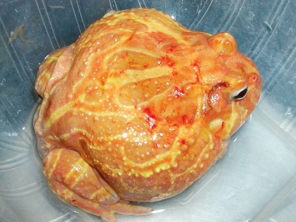
This pixie frog was bitten by a prey item. Feed your pixie frog pre-killed prey items when possible.
Fighting and bite wounds, rubbing of the nose on glass or screens or any other physical barriers can all be a source of potential infection. Covering glass walls with paint, etc., changing metal screen to soft nylon and separating large and small, or aggressive animals, may be necessary. Injuries should be addressed with first aid or even systemic antibiotics and pain medication as needed.
Amphibian Hypo/Hyperthermia
Hypothermia is generally not life threatening if caught early. The animal is slowly rewarmed back to its proper temperature. If exposed to cool temperatures for prolonged periods, immunosuppression is possible, as well as deleterious effects on the gastrointestinal tract. For instance, if an animal has taken a recent meal and then is exposed to temperatures that are too cool to support digestion, vomiting and intestinal upset can result.
DOUGLAS MADER, DVM, REPTILE MEDICINE AND SURGERY, 2ND EDITION
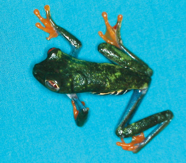
Starvation and dehydration are the most common nutritional problems encountered in captive frogs.
Hyperthermia can be lethal if not caught early. Signs include hyperactivity, incoordination, lethargy and, ultimately, death. As with hypothermia, the animal should be placed in fresh, chlorine-free water at the proper temperature to rapidly lower the body temperature. Animals that have been severely heat stressed may need veterinary intervention with fluids and corticosteroids.
Amphibian Toxicities
Amphibians are extremely sensitive to environmental toxins. This is likely due to their permeable skin and their high surface-area-to-body-weight ratio. Most all disinfectants are toxic to amphibians, including iodine, chlorhexidine, quaternary ammonium, chlorine and ammonia. Many of these compounds can absorb into plastic containers and subsequently leach out, even if no obvious disinfectant is visible, so it is best to use glass or stainless steel to contain them.
Signs of toxicity vary with species and toxin, and may include reddening of the skin, blood spots on the skin, increased mucus production from the skin, hyperactivity to lethargy, difficulty breathing, tremors and convulsions, paralysis, vomiting, diarrhea and death.
Pesticides and cigarette smoke have both been shown to be toxic to amphibians. It is best to keep amphibian areas smoke free.
Nutritional Diseases
Amphibian Nutritional Metabolic Bone Disease (NMBD)
More correctly knows as nutritional secondary hyperparathyroidism, this condition results from a triad of deficiencies involving UV light, available calcium in the diet and vitamin D3. It is second only to starvation or malnutrition in captive amphibians.
The most common sign of acute NMBD in amphibians is spastic tetany after strenuous movement. If not corrected, this will progress to more involved systemic changes, such as hydrocoelom (coelomic cavity filled with fluid) from gastrointestinal stasis (normal calcium levels are required for normal intestinal motility and digestion), spinal deformities, angular limb deformities and bowed mandibles that give the animal a grotesque “smile” appearance.
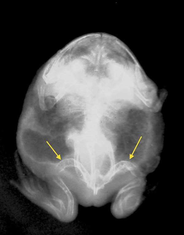
Douglas Mader, DVM, Reptile Medicine and Surgery, 2nd edition.
This White’s tree frog was suffering from NMBD. Note the disfigured thigh bones (yellow arrows) and the grossly mishappen jaw.
When caught in time, therapy for NMBD can be successful. Generally, the first step is correcting the underlying husbandry deficiencies (lighting, temperature, humidity, diet, etc.) and concurrently administering to the patient’s medical needs.
Animals showing signs of tetany will need systemic calcium supplementation. Animals not so affected will do well with oral calcium and vitamin D3 supplementation. Resolution of the NMBD may take weeks to months. Even with full correction of the deficiencies, permanent disfigurements may persist for the life of the animal.
Amphibian Hypovitaminosis A
The most common result of low vitamin A in the diet is a condition called short tongue syndrome, first described in the Wyoming toad (Anaxyrus [Bufo] baxteri). The low vitamin A levels create a condition called squamous metaplasia, which results in the animal’s inability to produce proper sticky mucus on the tongue. When a toad or frog tries to grasp prey, it doesn’t stick to the tongue when it is retracted into the mouth, and the prey gets away.
In addition to the effects on the tongue, hypovitaminosis A can affect the conjunctiva around the eyes (swelling), as well as the kidneys and the bladder (causing hydrocoelom).
Treatment involves correcting life-threatening conditions and supplementing vitamin A, as well as correcting the diet or any underlying cause.
Amphibian Obesity
Sit-and-wait predators, such as the horned, or Pac Man, frog, when overfed, can become obese in captivity. Although long-term effects of obesity in amphibians has not been studied, it can be assumed that detrimental effects can be seen, similar to that in other species.
Foreign Body Ingestion
Amphibians are keyed off to attack and eat items if they see movement. Because of its naturally sticky tongue, if the amphibian misses the prey and the tongue sticks to a cage item (like gravel or small rocks), these foreign materials may accidentally get ingested. While some animals are able to regurgitate the rocks out, this is not always the case. It is not uncommon to find rocks inside the stomach of larger, more aggressive frogs, such as the Pac Man frog. Treatment can be either reaching in with a forceps or endoscope to remove the items or, in some cases, surgery to take them out of the stomach.
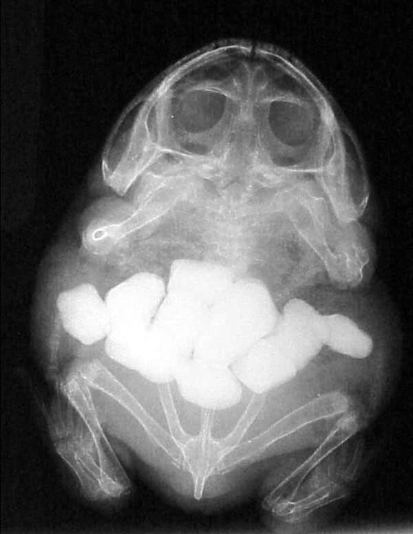
Douglas Mader, DVM
This X-ray shows multiple stones in a frog’s stomach. Surgery was needed to remove them and the patient did fine.
Amphibian Gastric Overload
Another issue involved with feeding is a condition called gastric overload or impaction (GOI). This results from overfeeding an amphibian too large a food item or excessive amounts in a short period of time. Although some anurans may regurgitate excessive meals, not all do.
Bloated, overfull stomachs can put pressure on the lungs, making it difficult for the animal to breathe. But more importantly, a large meal may not be digested properly, resulting in bacterial overgrowth and illness, and potentially death.
Treatment involves intervention, anything from stomach washes to actually going into the stomach with forceps or an endoscope and retrieving food items. In severe cases, surgical intervention may be needed. Gastric protectants and antibiotics may be required in some cases.
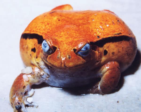
Amphibian Corneal Lipidosis
Whitish plaques, caused by deposition of cholesterol deposits in the cornea, are characteristic of this disease. High cholesterol levels in the blood, which are a result of diets high in fat, are generally believed to be the cause. Treatment is usually ineffective, but correction of the diet may prevent the condition from worsening.
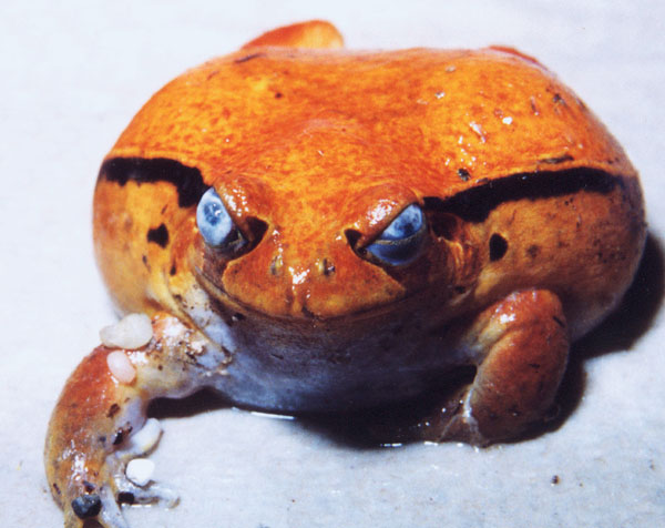
DOUGLAS MADER, DVM, REPTILE MEDICINE AND SURGERY, 2ND EDITION
Corneal lipidosis in a tomato frog.
Gout
Gout is the deposition of uric acid crystals in various locations within an amphibian’s body. It can be caused by several factors, including diets high in purines, dehydration, infection and kidney failure. The crystals can form in soft tissues, such as the liver and kidneys, but also form larger stones in the bladder. By the time it is diagnosed, it is often severe. Bladder stones are readily removed if the amphibian is in otherwise good condition.
Infectious Diseases
Amphibian Viruses
Amphibians are susceptible to numerous viral diseases. The most studied is the herpesvirus, which causes kidney tumors in the leopard frog. The virus affects the kidneys, causing hydrocoelom and hydrops. The animals begin to lose weight and ultimately die after spawning.
Iridoviruses (specifically the ranaviruses) have been the subject of much research in recent years. These viruses are responsible for die-offs in wild populations.
In general, there is little known about amphibian viruses, but more is being discovered because of the increased interest in disease in both captive and wild specimens.
Bacterial
Amphibians normally have bacteria that live within their bodies. Aeromonas, Pseudomonas, Proteus and E. coli are all considered normal flora. However, if the animal is immunosuppressed from underlying disease (e.g., iridovirus) or husbandry conditions (e.g., crowding) these commensals can become opportunistic pathogens, meaning that they can cause disease.
The classic red-leg disease, medically known as dermatosepticemia, is typically associated with Aeromonas hydrophila. In actuality, many different pathogens (bacteria, fungi, viruses) can all cause signs of dermatosepticemia. Clinical signs associated with this are usually rapid in onset and acutely lethal. Changes in the skin capillaries cause a red flush to the whitish areas on the legs. This can progress to subcutaneous hemorrhages and red-purple skin. Once this appears, death is usually imminent.
Treatment, which must be aggressive and immediate, targets the septicemia caused by the bacteria, but also adjunct therapy for chytrid fungus. Antibiotics, antifungals and fluids are generally required. Even with early, aggressive treatment, the likelihood of death is high.
Fungal
The most infamous fungus of amphibians is Batrachochytrium dendrobatidis, the agent for chytridiomycosis. Whereas most fungal infections are generally seen in immunocompromised amphibians, B. dendrobatidis can cause disease in healthy colonies and the wild.
Clinical signs of B. dendrobatidis vary from sudden death with no signs to slow onset, with reddening of the belly (similar to dermatosepticemia), lethargy and excessive skin shedding. If caught early, treatment can be effective.
Several other fungal organisms have been shown to cause disease in amphibians as well. Saprolegniasis produces cotton-like growths on the skin and in the mouths of aquatic amphibians. A pigmented fungi, chromomycosis, has been seen in various amphibians, where it produces light-tan to dark-gray nodules on the skin. Without treatment, death can result.
Given that most fungal diseases are the result of underlying poor environmental conditions and stress, correcting the deficiencies are mandatory for successful treatment and prevention of future disease.
Parasitic
Amphibians are host to various parasites, including protozoa, nematodes, trematodes and cestodes.
Amoebiasis and Entamoeba ranarum are common and can be treated if caught early. Nematodes, also known as roundworms, are the most common parasites seen in amphibians.
Rhabdias, known as the lungworm, can be found in small numbers in wild animals, and the combination of captivity and stress can result in heavy burdens and mortality. This is contagious to other amphibians, so affected animals must be quarantined and kept separate until properly treated.
Neoplasia
Amphibians are susceptible to neoplasia (cancer, tumors) just as seen in other animals. Cancer of the liver, kidneys, skin and gonads are the most common. Typical signs include hydrocoelom (coelomic cavity filled with fluid). Diagnosis and treatment varies, but in general, the prognosis is guarded to grave.
Summary
Amphibians can make for excellent pets or display animals. A thorough understanding of their biology and natural history prior to acquiring them is paramount to maintaining a healthy population. When disease occurs, early recognition and intervention will maximize the chances for a favorable outcome.
This article is dedicated to Kevin Wright, DVM, DABVP (Reptile/Amphibian), 9/27/62 — 9/26/13.
Douglas R. MADER, MS, DVM, DABVP (REPTILE/AMPHIBIAN), is a graduate of the University of California, Davis. He owns the Marathon Veterinary Hospital in the Conch Republic, and is a world-renowned lecturer, author and editor. He sits on the review boards of several scientific and veterinary journals.

