Stick tail in leopard geckos is often caused by an intestinal infection.
“Stick tail” is a lay term for weight loss in leopard geckos and fat-tail geckos. Often, it is caused by an intestinal infection by Cryptosporidium varanae (formerly Cryptosporidium saurophilum). Other common causes are gastrointestinal infections of flagellated protozoa or Gram-negative bacteria. Internal abscesses or granulomas are common. As-yet-undescribed viruses may play a role. There are many other conditions that can cause weight loss and stick tail.
Stick Tail Symptoms/Signs
Weight loss and shrinking of the tail fat deposits until the tail is little more than skin covering the caudal vertebrae. Some geckos may have white spots on the liver that are visible through the belly skin. Stick tail is often accompanied by diarrhea, poor appetite or anorexia, hiding, and spending time in the coolest parts of the enclosure. It often affects more than one gecko per cage and may affect multiple cages in a breeding colony.

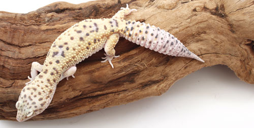
Photo credit: Gina Cioli/I-5 Studio
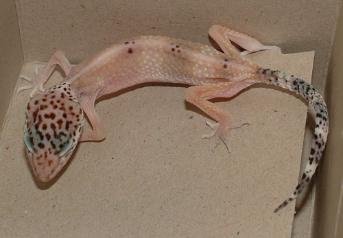
Compare the top photo of a healthy leopard gecko’s tail with the photo beneath it of a gecko exhibiting stick tail.

Photo by Kevin Wright
An adult, malnourished leopard gecko exhibiting stick tail.
Species at Risk
Leopard geckos and fat-tail geckos, though many other lizards may have similar underlying causes of weight loss.
Predisposing Captive Conditions and/or Other Factors
Any suboptimal husbandry practice may cause immunosuppression and increase susceptibility to infectious diseases. Among the more common factors are cool temperatures (e.g., those that are below the Preferred Operating Temperature Zone), crowding, unsanitary conditions (allowing droppings, uneaten food and other waste to build up in a cage), poor hygiene (transferring waste and contaminated material between cages), skipping or insufficient length of time spent in quarantine, prolonged shipping time and insufficient vitamin A in the diet.
Diagnostic Tests a Veterinarian May Recommend
Fecal parasite tests will detect flagellated protozoa, coccidia and many other intestinal parasites. A PCR test will detect Cryptosporidium varanae. Bacterial cultures of the cloaca may reveal Salmonella or other important bacterial pathogens. Adult leopard and fat-tail geckos are large enough for bloodwork to detect kidney, liver and other internal diseases.
Safe Practices/Prevention
Isolate: Affected geckos should be isolated. This helps prevent further spread of the disease and may remove them from stressful competition with cagemates.
If an infection is confirmed, any geckos that were cagemates or any geckos that may have otherwise been exposed to the disease should be isolated. Screening for that particular pathogen is recommended. If tests do not detect infection and the geckos appear to be healthy and maintaining or gaining weight over the next 45 days, they may be considered at low risk of carrying the infection. Any geckos that test positive should be treated. Any geckos that are losing weight or otherwise appear unhealthy should be kept in isolation until an underlying cause is determined.
Quarantine: All incoming geckos should be quarantined to prevent introduction of Cryptosporidium, flagellated protozoa and other infectious diseases. Recommended screening tests for geckos are fecal parasite examinations at three different times and one Cryptosporidium PCR test. These should show no significant parasites for a gecko to be considered for release. As a general rule, a gecko should be eating well, maintaining or gaining weight, free from common detectable infectious diseases and appear healthy for one month before it is released from quarantine or isolation.
Proper hygiene and sanitation: Organic material should be thrown out because it cannot be easily disinfected. A sweater box works well as a simple, easy-to-clean hospital cage when furnished with paper towel substrate, a plastic hide box, and plastic food and water dishes. Spot-clean the cage daily to remove droppings, uneaten food, shed skin and other waste. Deep-clean the cage and all furnishings weekly with warm soapy water, followed by a 15- to 30-minute soak in a disinfectant such as dilute chlorine bleach. Rinse well so that no disinfectant remains on the cage or any of the cage furniture. Clean any tools used in or around the cage and disinfect as mentioned.
Handle infected geckos and cages only after you have completed all tasks required for any other reptiles you may keep. If Cryptosporidium is detected, full-strength household ammonia (5- to 10-percent ammonia) should be used as a disinfectant. Do not mix ammonia and bleach, as this produces a gas that is quickly fatal to people and animals.
Treatments a Veterinarian Would Likely Recommend
Any medication or other procedures recommended will depend on the underlying cause of the gecko’s weight loss. Paromomycin is a medication that helps control many cases of Cryptosporidium varanae and may be continued as a lifelong treatment. Metronidazole and ronidazole are often prescribed for flagellated protozoa and certain kinds of bacterial infections. Trimethoprim-sulfa, amikacin, enrofloxacin or other antibiotics may be prescribed for Gram-negative bacterial infections.
An affected lizard’s intestine may be damaged and not absorbing water and other nutrients properly. Daily soaking in chin-deep, cage-temperature water may be needed to help keep a gecko well-hydrated, or it may need to receive injectable fluids. Liquid diets such as LaFeber’s Emeraid for Carnivores or Oxbow’s Carnivore Critical Care may be given orally to help a gecko regain weight. Liquid calcium may be needed if the gecko is at risk of nutritional secondary hyperparathyroidism.
It is important to work with your veterinarian to get as accurate a diagnosis as is practical for your stick tail gecko. Otherwise, you could waste time with a current treatment, prolonging your gecko’s illness and putting your other reptile pets at risk.
Prognosis
Prognosis varies depending on the underlying cause. Simple gastrointestinal infections with flagellated protozoa often respond well to treatment. Internal abscesses or granulomas have a poor-to-grave outlook and euthanasia is recommended. If a gecko infected with Cryptosporidium varanae does not show weight gain, increased activity, and a willingness to eat on its own within three weeks of treatment, its outlook is poor and euthanasia should be considered. The more information your veterinarian has, the better he or she can advise you as to your gecko’s chances.
Can Humans Catch It?
Most causes of stick tail are not zoonotic. However, Salmonella has been cultured from ill geckos and should always be considered. Always take proper precautions, such as washing your hands with warm soapy water after handling a sick gecko.


