Ailments of tortoises in captivity and potential treatments.
Tortoises are popular as reptile pets. They are interesting and will thrive and give the hobbyist many decades of pet-owning pleasure (many species can live 60 years or more!) as long as they are properly maintained and cared for. In this article I will discuss three common ailments that may be encountered in pet tortoises and what you should look out for.
1. Tortoise Respiratory Infections
Respiratory system infections are common in pet tortoises, especially in wild-caught individuals sold in the pet trade. Being shipped from their native lands and held in less-than-desirable circumstances causes a great deal of stress on the tortoise. Add to this that often the poor village people collecting them may lack the means (heated houses, running water, available food, sanitation, etc) to house them properly before shipment ever takes place. Many times tortoises are held in great numbers in small un-heated enclosures for weeks to months, with little food or water. Reptile brokers arrive every few weeks to months to buy these poor creatures, and then ship them overseas, again, often in crowded, unsanitary shipping boxes or crates.
By the time these tortoises arrive at their destination, they commonly show signs of lethargy, ocular and nasal discharge, weight loss (a healthy tortoise should feel heavy for its size, like it’s shell is filled with water), with increased respiratory rates and wheezing. These ill tortoises will often refuse food despite being given proper care and an environment.
Captive-raised tortoises can also show these signs if they have not been properly cared for or fed correctly. This is less common than in wild-caught individuals, but nevertheless it is seen. Be careful who you obtain a pet tortoise from. Ask questions as to when it was born, past health history, diet fed, etc.
Respiratory infections (RI) are best diagnosed and treated by a veterinarian who works with reptiles. A thorough physical examination and detailed history from you the owner (asking where was the tortoise purchased, captive raised or wild caught, husbandry, diet, etc) is where the veterinarian will start. Radiographs of the lungs and a blood panel may also be necessary to fully access how ill the tortoise is and to give a prognosis for treatment.

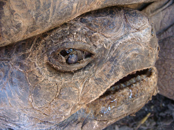
Photo by Kenneth A. Harkewicz, VMD
Aldabra tortoise with conjuctivitus due to a respiratory infection.
Treatment usually entails a course or antibiotics, often for several weeks to months (depending on the severity of the RI). These can be given orally, but give that these tortoises often are not eating and have strong jaws and withdraw into the shell even when ill, the treatment is often given via injections. Most veterinarians will show owners how to administer the medication (it’s not hard once learned). Nasal and ocular drops are also sometimes administered along with the antibiotics to help the turtle smell and see its food better.
In severe cases, a feeding tube is sometimes placed surgically through the neck of the turtle so it can be fed a gruel to maintain its strength until eating on its own. These feeding tubes may be in place many weeks to months.
Keeping the tortoise warm during treatment is also a necessity. The veterinarian may suggest keeping them indoors under heat and full-spectrum lighting until well. This may be a problem with large species, such as Sulcata tortoises, which can weigh up to 125 pounds or more. Something to consider when purchasing one of these as a cute little hatchling!

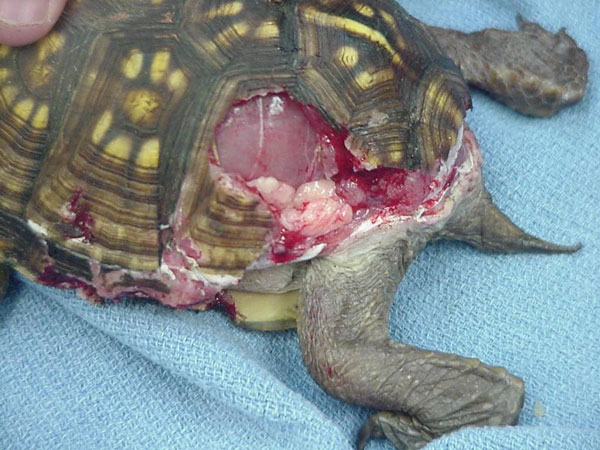
Photo by Kenneth A. Harkewicz, VMD
This chelonian was attacked by a dog.
Respiratory infections can be prevented in several ways. Always buying captive-raised rather than wild-caught tortoises increases your chance of obtaining a health(ier) tortoise from the start. It’s not a guarantee, but your chances are much better.
Keeping the tortoise in a proper enclosure with adequate heat, offering full-spectrum lighting and feeding a proper diet makes for a healthy tortoise. Each tortoise species has its own set requirements for proper care, so being informed of your pet’s needs is important. REPTILES magazine, books on your tortoise species, reptile clubs and online forums, such as those found on ReptileChannel.com, are all are good places to obtain information.
Finally, having your veterinarian examine the tortoise at the first sign of a respiratory infection really helps in curing this condition. Waiting to see if the pet will get well on its own, or trying home/over-the-counter products, only prolongs the infection and lessens the chance for a full recovery. Don’t delay treatments!
2. Metabolic Bone Disease (Soft/Deformed Shells) in Tortoise Shells
Metabolic bone disease (MBD), also known as nutritional secondary hyperparathyroidism (NSHP), can be a common aliment of pet tortoises. This is due to a lack of calcium in the diet or a problem absorbing dietary calcium.
Tortoises need exposure to ultraviolet radiation A and B (UVA/B) in order to manufacture vitamin D3 from vitamin D2. Vitamin D3 is necessary for calcium in the diet to be absorbed through the gastrointestinal tract. In the wild, the sun provides ultraviolet light. Wild tortoises bask 8 to 14 hours/day and receive the necessary UVA/B rays in order to absorb calcium from food. Sunlight that shines through a glass or plastic window or cage is blocked from providing the necessary radiation.
Tortoises fed a diet low in calcium, or those who cannot absorb calcium well, usually first show a soft shell, especially if they are a juvenile. Baby tortoises naturally have a semi-soft shell after hatching, but it usually hardeners by 6 to 8 months of age. In calcium-deficient tortoises, the tortoise grows, but the shell does not. This causes the shell to become deformed and often the tortoise looks too big for its shell. It become weak, and eventually has trouble walking, due to soft bones. The soft bones can easily fracture, causing further problems. If untreated, the tortoise will eventual die. This is often a slow, painful death.
Metabolic bone disease can be prevented in several ways. First, a full-spectrum light source is needed in indoor enclosures. Those tortoises kept in outdoor pens get full sunlight as long as the pen/cage is placed where the sun shines. If they are housed indoors during the winter, provide the right lights. These can be incandescent or fluorescent, and they should be placed onto of the tank without any glass or plastic blocking it from the tortoise (screening is OK). The light should be as close as possible, between 12 and 18 inches away from the turtle. You want it close, but not close enough to where thermal burns can occur. Leave these full-spectrum bulbs on 12 to 14 hours/day, and change them every six to 10 months; their UVB output lessens over time.
The diet should be on rich in calcium. Beet tops and dark, leafy greens are an excellent source of calcium (iceberg or Romaine lettuce are not). A powdered vitamin/mineral supplement can be dusted over the food to ensure added calcium. These can be purchased at many pet-supply stores and online.
Treatment should first be a trip to your herp veterinarian. He/she can assess the degree and severity of the MBD, usually by using a radiograph to see if pathologic fractures have occurred. A blood panel is sometimes run, making sure other body systems have not been affected.
Husbandry and diet need to be corrected if inadequate. Sometimes oral liquid calcium is prescribed in addition to the above recommendations.
Tortoises severely malformed from MBD may never look normal. The misshapen shell may never appear like that of a healthy tortoise, especially if this has been a long-term condition.
3. Tortoise Prolapses
The cloaca in reptiles is the opening under the tail for the digestive tract, the reproductive system and the bladder. All three empty out the same cloacal opening. Tortoises will sometimes prolapse tissues/objections out through this opening.
Tortoises produce uric acid in their kidneys to expel fluids as urine. Unlike mammals that produce water-soluble urea, the uric acid that reptiles make is not soluble in water. It can look like milk in the case of diluted urine, or hard chalk in concentrated urine.
Due to dehydration or a poor diet (one high in oxalates found in some plants/vegetables), a hard “stone” of the material may be produced in the bladder. The tortoise attempts to pass this stone by pushing its abdominal muscles against the bladder. If the stone is too large, or the tortoise is too weak, organs rather than the stone may be passed out through the cloaca. In males, the phallus may be pushed out. Male tortoises do not have a true penis. Rather they have a phallus. This is a spear-shaped organ through which sperm pass when mating (urine is emptied from the bladder into the cloaca). If the phallus becomes dry or damaged when exposed to the environment, it may not be able to place itself back into the body cavity. If the animal continues to strain and is in discomfort, this becomes a medical emergency. Take the tortoise to a veterinarian ASAP!

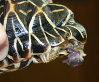
Photo by Kenneth A. Harkewicz, VMD
Star tortoise with phallus prolapse.
The veterinarian will first try to gently place the organ back inside. Applying a hypertonic sugar solution (i.e. 50-percent-dextrose solution) will draw fluid out of the swollen phallus through osmosis to shrink it. The organ is then lubricated (K/Y Jelly, a water-based lubricant is often used) and gently pushed back into the cloaca. A purse-string suture is then usually placed into the skin of the external cloacal opening to prevent it from coming out again while the tortoise recovers. The sutures are left in place for five to seven days. This usually works in cases that are seen early after prolapse. If the organ is too dry, or the attempt to replace is not successful, the phallus may need to be amputated under sedation/general anesthesia. The tortoise is fine; it just cannot reproduce.
It is important to address why the organ prolapsed and correct this. If it was due to a urate stone, diet and hydration needs to be corrected. Large stones sometimes need to be removed surgically through the plastron (bottom shell). This is an expensive and involved surgery. Small stones are usually passed with soaking/hydration.
Male tortoises can sometimes bruise the phallus during mating, which can cause a prolapse. If this happens repeatedly, it’s best to separate the sexes.
Tortoise Ailment Prevention through Proper Husbandry
These three tortoise aliments can all usually be prevented through proper husbandry, diet and care. Become familiar with your tortoise’s needs, and it will give you many years of enjoyment.
Kenneth A. Harkewicz, VMD Berkeley Dog and Cat Hospital, Berkeley, Calif., is an exotic animal veterinarian, practicing in the San Francisco Bay area, and the staff veterinarian of the East Bay Vivarium, one of the nation’s largest retail herptile stores. He is also the current president of the Association of Reptilian and Amphibian Veterinarians (ARAV).


